
Multimodal Image Analysis
(マルチモーダル画像解析)
Development of image correction method for micro-CT images
|
Takayuki Okamoto,Daiki Matsumoto |
| Micro-computed tomography (micro-CT) is an X-ray imaging device that enables 3D imaging of a micron order's inner structures. Micro-CT has been generally used as a non-destructive testing in the industrial field, but the imaging of pathological specimens can provide new pathological information that cannot be obtained by conventional pathological diagnosis. However, in the case of pathological specimens imaging, there are problems such as the long scanning duration to acquire high-quality images and the influence of noise. We are developing high-speed imaging and image correction methods for pathological diagnosis using micro-CT. |
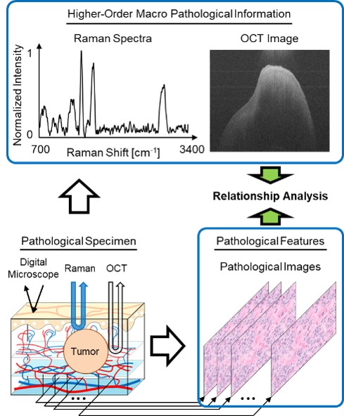
Higher-Order macro pathological information analysis by optical measurements
|
Kotaro Takahashi |
| In our laboratory, in order to obtain and analyze higher-order macro pathological information, we are working on constructing optical measurement systems using a spectroscopic camera, Raman spectroscopy, optical coherence tomography (OCT), and digital microscope, and developing image analysis methods. In addition, for the sake of obtaining information on structures and compositions under the mucosa, we aim to construct a mechanism to control the wavelength and spatial pattern of light irradiation. |
|