

Registration of multimodal image
(マルチモーダル画像のレジストレーション)
Relationship analysis between acoustic features by microscopy and pathological characteristics
|
Daiki Matsumoto, Shu Kashio |
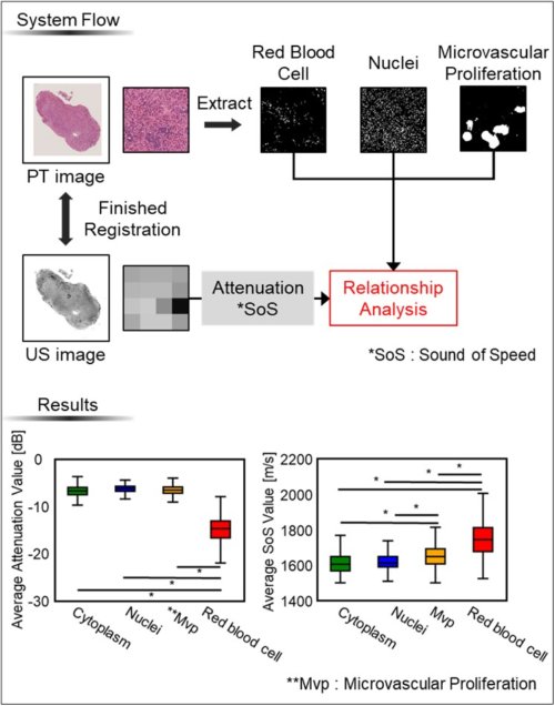
| In the case of brain tumor removal, the grade of the tumor removed intraoperatively is judged qualitatively by its color and hardness. In addition, the pathological analysis to make a final diagnosis of the type and grade of the tumor after surgery requires more time. Therefore, we focused on the ultrasound system, which is a modality with information correlated with hardness, as a quantitative and immediate tumor determination system. Intraoperative ultrasound diagnosis and pathological features are difficult to combine because of the difference in resolution, so we aim to analyze the relationship between ultrasound measurements and pathological features. |
 |
Registration of multimodal image
|
Takuya Tanaka,Shu Kashio, Noriaki Hashimoto |
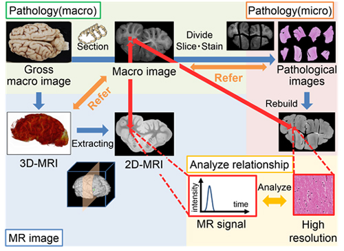
| In the diagnosis and treatment of head disease, diagnostic imaging is valid. However, to get pathological information from MR image, it needs to analyze relationship between MR image and pathological images in the same region because it needs pathological information of target tissue for determining the method of treatment. However, it couldn’t be compared to MR image directly because the pathological images is divided and deformed through making pathology specimens. This study was made to register pathological images and MR image via optical macro images taken before making the tissue specimen. Referring to optical macro images, we deformed and corrected pathological images and searched a single MR slice matched to an optical macro image from 3D-MR images. Finally, we registered the corrected pathological images and extracted MR slice. As a result, we could compare pathological images to MR image of the identical area. |
 |