

Knee Joint Image Analysis
(膝関節画像解析)
Registration and segmentation of multi-contrast MR images for knee cartilage evaluation
Aizimu Tuerxun, Toru Tanaka
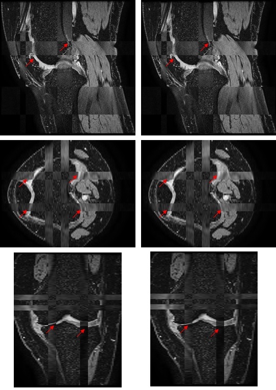
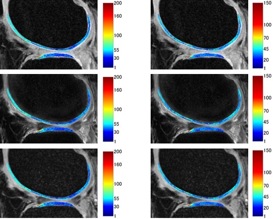
| T1ρ and T2 relaxation time evaluation of multi-contrast magnetic resonance images for evaluation Magnetic resonance imaging is a suitable, noninvasive method for the quantitative evaluation of articular cartilage of patients with knee osteoarthritis. The T1ρ and T2 relaxation times are two advanced magnetic resonance based cartilage evaluation methods. Our research is focused on multi-contrast magnetic resonance images for T1ρ and T2 relaxation time evaluation. Slight misalignment of each bone region is separately corrected by a rigid transform. The cartilage region was segmented using multi-contrast magnetic resonance images. We used in-vivo magnetic resonance image data from patients to evaluate our method and thereby, confirmed the accuracy of the proposed method. |
 |
 |