
Imaging and analysis of microcirculation
(微小循環のイメージングと解析)
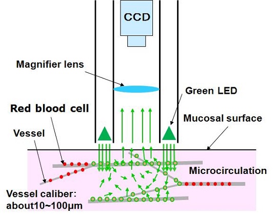
Microcirculation is the small blood vessels whose diameter is less than 100 μm and is found in all parts of the body. Its functions areis gas and nutrient exchange between the blood and the tissues. It ensures adequate immunological functions. The microcirculation is markedly altered in sepsis, that these alterations are more severe in nonsurvivors than in survivors. So, the monitoring of microcirculation is needed for understanding the biological condition.Development of Sidestream dark-field (SDF) imaging
|
Sinko Ri, Tomohiro Kurata, Minori Takahashi, Kodai Nihei |
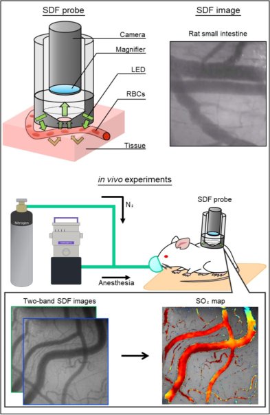
| Sidestream dark-field (SDF) imaging was developed as an imaging system to observe microcirculation noninvasively. We have developed an SDF probe using three color LEDs and it produces images of three wavelength by switching the LED light sources. As a result, we verify that SDF imaging allows shooting blood flow of red blood cells (RBCs) at organs of the pig and sublingual of the human. By utilizing the difference in the spectral characteristics of oxygenated and de-oxygenated hemoglobin in the RBCs from the spectral image, we are aiming to establish a blood oxygen saturation measurement technique. Furthermore, to evaluate quantitatively the blood flow by analyzing a moving image, we are aiming to monitor the state of health. |
 |
Oxygen Saturation Mapping of Microcirculation Using Sidestream Dark-Field Imaging
|
Ryohei Hashimoto |
| Sidestream dark-field (SDF) imaging is a noninvasive and clinically applicable technique to observe microcirculation. So, we have developed an SO2 estimation method based on SDF imaging. Our proposed method utilizes two-wavelength oximetry based on the Beer–Lambert law. Then, we conducted the in vivo experiment observing the changes in SO2 of microcirculation using SDF imaging. |
 |