

Visualization and Analysis of Motion of Thoracoabdominal Organs
(胸腹部の呼吸性運動の可視化と解析)
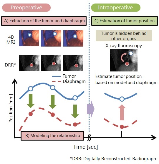
Lung tumor tracking based on thoracic 4D-MR image for radiotherapy
|
Shuhei Isa(graduate) |
| The radiotherapy is employed for cancer treatment. It is able to destroy tumor mass by exposing to radiation. However if the normal tissue is exposed to radiation, it is also destroyed. Therefore, in radiotherapy of the thoracic region, it is important to grasp the tumor motion due to respiration in order to avoid unnecessary exposure to healthy tissues. Generally, the tumor is tracked using X-ray fluoroscopy. However, the tumor is sometimes hidden behind other organs and direct tracking is very difficult. In such case, diaphragm location can be used as a surrogate to estimate the tumor location. In this study, we are developing an estimation scheme of tumor location with preoperative 4D (spatiotemporal)-MR image. In this scheme, we first model the relationship between the tumor location and feature points location on the diaphragm based on thoracic 4D-MR image. During the treatment, tumor location is estimated from the diaphragm in the X-ray fluoroscopy. |
 |
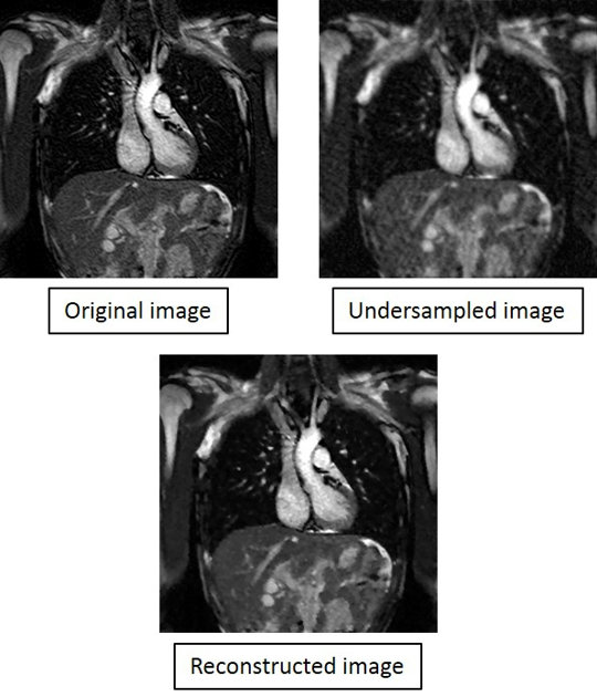
Reconstruction method by using sparse
|
Yukinojo Kitakami(graduate) |
| 4D-MRI can visualize and quantify the three-dimensional dynamics of the thoracoabdominal respiratory movement and allows us various applications. However, one of the problems is long data collection time, approximately 30 minutes. In this study, we assume to reduce the number of the encoding in the k- space to shorten the data collection time. However, images are degraded by this reduction. Therefore we used the reconstruction technique that used sparse modeling such as compressed sensing. |
 |