
X-ray Imaging Application
(X線画像応用)
IVR Assisting System
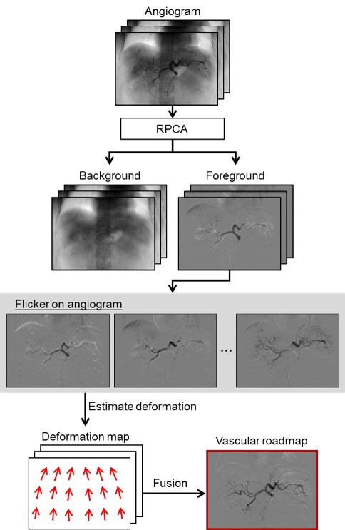
| Interventional radiology (IVR) mainly refers to catheterization, which is less invasive than open surgery. In the procedure, a position of the surgical tool is determined by using X-rays to see inside the body. However, vascular vessels do not appear on X-ray fluoroscopy. Therefore, the method called Digital Subtraction Angiography (DSA) is used to remove background information such as bone and to enhance the vascular vessels. Patients need to hold their breath during DSA, and that is a burden to them. Then, we aim to develop a method of vessel enhancement by motion picture processing without DSA. |
Vascular Enhancement of from Free-breathing Angiography and Generation of Vascular Roadmaps
|
Morio Kawabe, Yuri Kokura |
| Robust principal component analysis (RPCA) is used to separate contrast components, such as vascular components, in angiograms taken under naturally breathing environment. We also focus on reducing the processing time, which is a problem for the conventional RPCA. To solve the problem of flickering contrast, we are generating a vascular roadmap by fusing multiple angiograms. We aim to contribute to the surgical field visibility in IVR. |
 |
X-ray tomosynthesis from images obtained in free imaging geometry
|
Kohei Sato |
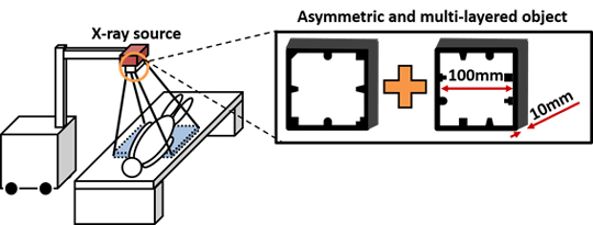
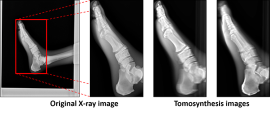
| Tomosynthesis is attracting attention as a low dose tomography technology compared with the X-ray CT. However, conventional tomosynthesis imaging devices as well as X-ray CT devices are still large and stationary. Furthermore, there is a limitation in the working range of X-ray source during the image acquisition. In this study, we propose to use a portable X-ray device that is widely used for round and emergency medicine. Tomosynthesis using a portable X-ray device requires the calibration of the geometric relationship between the X-ray source and the detector at each image acquisition. Such tomosynthesis with a free imaging geometry also requires an iterative reconstruction. We present an image processing-based calibration method using an asymmetric and multi-layered calibration object attached to the X-ray source housing. We then utilize an iterative image reconstruction method. |
|
|
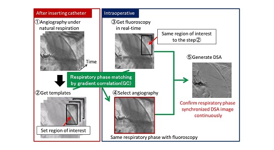
Respiratory synchronized roadmap by use of respiratory phase matching between angiography obtained after inserting catheter and intraoperative fluoroscopy
|
Yuya Takano |
| Intervention with a catheter is conducted under X-ray fluoroscopy. In this intervention, angiography and digital subtraction angiography (DSA) are conducted to enhance blood vessels. However, displacement and transformation of vessels caused by respiration are not considered in DSA because an image for only one phase can be acquired during breath holding. In this study, we propose a new method for generating a vessel roadmap corresponding to all respiratory phases and synchronizing with fluoroscopy by using a pattern matching between angiography after inserting catheter under natural respiration and intraoperative fluoroscopy. |
|
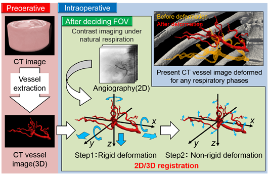
Three-dimensional respiratory deformation processing for CT vessel image using angiographic images during a respiratory cycle
|
Shohei Suganuma |
| In interventional radiology, physician inserts a catheter into blood vessels and forwards it to a target region under fluoroscopy monitoring to perform the diagnosis and treatment. However, since the vessels are deformed due to respiratory motion, the guidance of catheter is often difficult especially for novice physicians. On the other hand, CT image is also used for understanding the three-dimensional structure of blood vessels in addition to fluoroscopy. However, such a CT image does not have the respiratory motion. In this study, we propose to generate CT images with respiratory deformation from the 2D/3D registration between a preoperative CT image and continuous angiographic images acquired intraoperatively under natural respiration. |
|