Ultrasonics Laboratory for Tissue Characterization (UTLC)
ULTC is a laboratory operationg at the ultrasonic group of the Centrer for Frontier Medical Engineering Center, Chiba University. ULTC is conducting researches on ultrasound in medicine, such as acoustic measurement and development of diagnostic/treatment support technology under the leadership of Professor Tadashi Yamaguchi. The main research topics are the ultrasonic quantitative diagnosis includes the standardization and the tissue property evaluation. The education and researches are being promoted in collaboration with multiple institutions and companies with the motto "Understanding the essential relationship between the physical/chemical phenomena and the wave phenomenon to be measured".
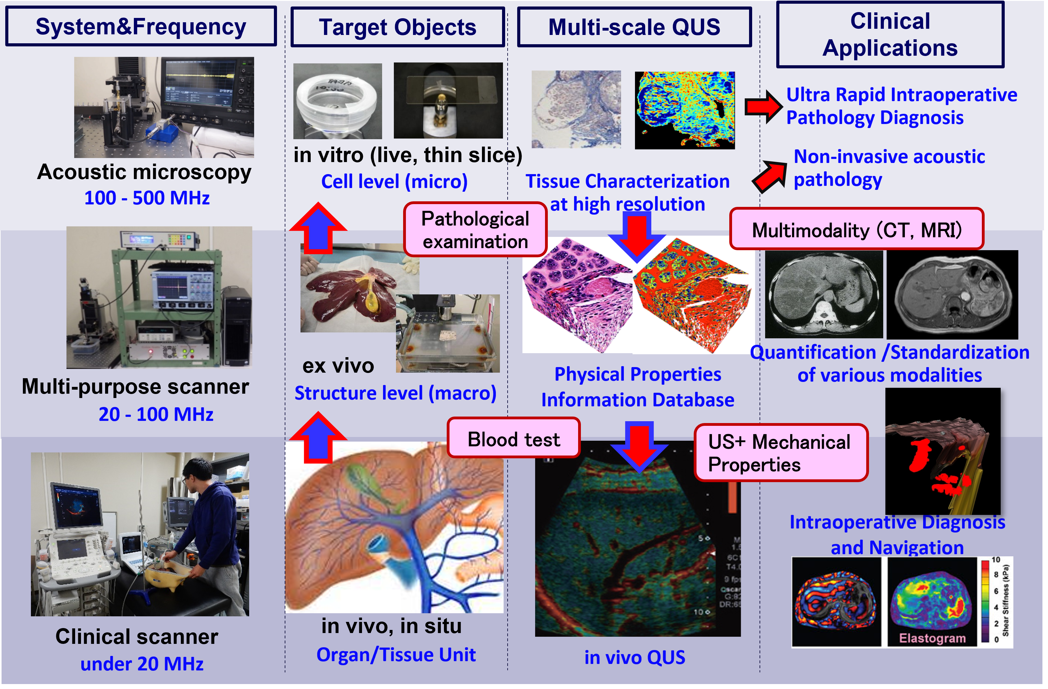
Fig. Understanding and quantitative indexing of biophysical properties at multi-scale and multi-modality.
Acoustic Properties Measurement by Ultra-High Frequency Ultrasound
- Measurement of the speed of sound, attenuation and acoustic impedance by 1 MHz-250 MHz Ultrasound
- Verification of the relationship between tissue composition/structure and acoustical characteristics.
Fig. Speed of sound of fibrotic rat liver measured by 250 MHz ultrasound (left). HE staind histology of same tissue (right).
Tissue Characterization by Multi-Band Frequency Ultrasound
- Detection of small target tissues from echo signal/image of each organ.
- Quantitative diagnosis of progressive diseases by statistical analysys of echo signal.
- Determination of metastasis by high-frequency ultrasound.
Fig. Fiber structures in serious chronic hepatitis human liver.
Fig. Estimation result of scatterer density in metastatic human lymph node.
3D Fusuin Imaging for Surgery Assistance
- Visualization of target tissues in each organ.
- Segmentation/Registration of targrt tissues in US, CT and Oputical image.
- Muiti-modality fusion imaging.
Fig. 3D fusion imaging of ultrasonic imaeg and laparoscopic image (left). visualization of blood vessels in liver (right).
Facilities
- Clinical ultrasound scanners (Canon, GE, Siemens, Honda Elec.)
- Research Ultrasound System (Verasonics)
- Ultrasound bio microscopies (Honda elec, UITC original)
- Multi-purpose ultrasound sccaner (UITC original)
- Single-element transducers ranging from 0.5 to 500 MHz
- Optical benches with a 3-axis computer-controlled motion system.
- A vacuum chamber and a churning machine for making phantom
- A surgical bench for small animal
- Large set of phantoms for accuracy evaluation of ultrasound scanner and transducer
- X-Ray CT and micro X-ray CT for animal experiments (owned by CFME)
- Open MRI for animal experiments (owned by CFME)
- MRI microscopy (owned by CFME)
- Breeding/experiment rooms for large/small animals (owned by CFME)
- Cell laboratory including fluorescence microscope (owned by CFME)