※以下のとおり開催しました。多くのご参加ありがとうございました※
January 17 (Wed), 2018
International Young Researchers' Workshop on Multimodal Medical Engineering 2018-若手研究者ワークショップ- 詳細はこちらから
January 18 (Thu), 2018
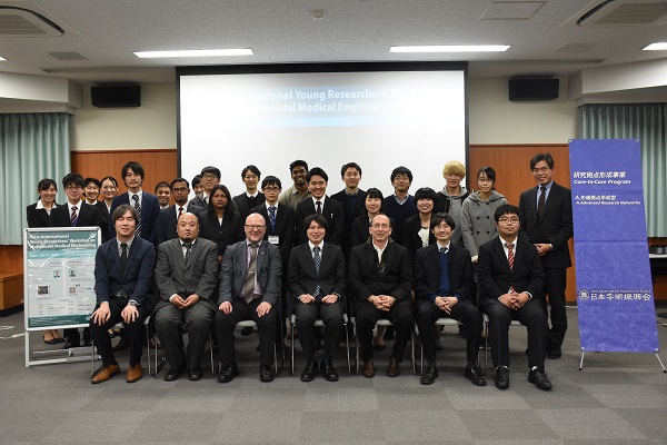
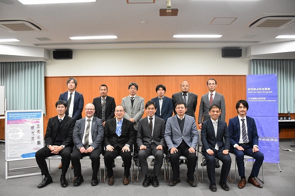
Second International Symposium on Multimodal Medical Engineering (MME)
- 第2回マルチモーダル計測医工学国際シンポジウム-
Venue: Chiba University, Engineering Research Building 2 Conference Room
(千葉大学西千葉キャンパス工学系総合研究棟2 2階コンファレンスルーム)
〒263-8522 1-33, Yayoi-cho, Inage-ku, Chiba 263-8522, Japan
千葉県千葉市稲毛区弥生町1-33
※ Download Leaflet (ポスターのダウンロードはこちらから)
Organized by:
“Multimodal Medical Engineering project” in Strategic Priority Research
Promotion Program, Chiba University
Supported by:
1. Institute for Global Prominent Research, Chiba University
Website: http://www.cfme.chiba-u.jp/~haneishi/mmme/sympo.html
2. JSPS
Program
Opening remarks 10:00-10:15
・ Takeshi Tokuhisa, President, Chiba University
・ Minoru Seki, Executive Director, Chiba University
Overview of MME10:15-10:30
・ Hideaki Haneishi (Chiba University)
Session 1: Outcome of the MME project 10:30-11:30 20min x 3
Multi-scale Biomechanical Modeling in the Cardiovascular System and Clinical
Applications
・Hao Liu, CFME, Chiba University
Biological tissue characterizations by electromagnetic field
・Kazuyuki Saito, CFME, Chiba University
Combination of contrast-enhanced ultrasonography and near-infrared
fluorescence using microbubbles with indocyanine green derivative
・Kenji Yoshida, CFME, Chiba University
Poster session 11:30-12:30 (Papers from CU)
1 Fluorescence imaging and spectral estimation for tongue coating measurement
Yudai Ota, Toshiya Nakaguchi, Vladmir Bochko, Pauli Falt, Markku Hauta-Kasari
2 Tongue Color Spatial Analysis based on Tongue Shape Normalization
Kazunari Murai, Toshiya Nakaguchi, Akira Morita, Takao Namiki
3 Endoscopic liver tracking for Water-Filled Laparoendoscopic Surgery (WaFLES)
Mika Kontto, Ryoichi Nakamura
4 Quantitative evaluation of endoscopic intestinal suture training usingtime
series
process analysis
Seiya Aoki, Naoki Fukatsu, Tomoko Yamaguchi, Munenori Uemura, Makoto
Hashizume, Ryoichi Nakamura
5 Development of integrated master manipulator with both forceps controller
and surgical workspace creator for single port WaFLES
Yuma Shimura,Kazuya Kawamura
6 Basic evaluation of power transmission using metal wire and mechanical
links for
forceps manipulator in single port WaFLES
Yusuke Tagata,Kazuya Kawamura
7 Development of a MR elastography system using low static magnetic field
open-MRI
Hiroki Yoshida, Mikio Suga
8 Triple-gamma imaging simulation of a novel Compton-PET system
Okumura Yusuke, Eiji Yoshida, Hideaki Tashima, Mikio Suga, Naoki Kawachi,
Katia Parodi, Taiga Yamaya
9 Scattering property estimation of beginning fatty liver disease
Kazuki Tamura, Kenji Yoshida, Hiroyuki Hachiya, Tadashi Yamaguchi
10 Evaluation of diffused liver disease by means of microscopic acoustic
characteristics
analysis with ultra-high frequency ultrasound
Kazuyo Ito, Takuya Ogawa, Tamaki Honda, Kenji Yoshida, Hitoshi Maruyama,
Tadashi Yamaguchi
11 Developmetn of a multimodal drug delivery system for reatments of malignant
diseases
Yiting Zhang, Masahiko Ebata, Taro Toyota, Hideki Hayashi
12 Development of a microbubble/near-infrared fluorescence dual imaging
contrast
agent for laparoscopic surgeries
Masahiko Ebata, Kenji Yoshida, Yiting Zhang, Taro Toyota, Hideki Hayashi
13 Performance evaluation of microwave surgical device by microscopic images
of
cells
Ryo Manago, Kazuyuki Saito
14 SAR evaluations of human body close to Wi-Fi base station
Kosuke Nishino, Kazuyuki Saito
Lunch 12:30-13:30
Session 2: Keynote Lectures 13:30-15:00 30 min x 3
High Frame Rate Ultrasound Imaging and Its Application to Cardiovascular
Functional Imaging
・ Hideyuki Hasegawa (Ultrasound), University of Toyama
Physiological imaging of in vivo biological tissues based on diffuse reflectance
spectroscopy
・ Izumi Nishidate (Optics), Tokyo University of Agriculture and Technology
Simultaneous PET and MRI imaging: how can the systems be affordable?
・ Taiga Yamaya (PET), National Institute of Radiological Sciences, QST.
Session 3: International Network of Multi-modal Medical Engineering for
PrecisionMedicine (JSPS Core-to-Core Program Session) 15:30-17:40
・ Hideaki Haneishi, Overview of Activities in FY2017, 10min.
Spectral based eye fundus imaging
・ Markku Hauta-Kasari (School of Computing, University of Eastern Finland,
Finland) 30 min
Initialization of active contours and level set method for segmentation
of ultrasound images of breast cancer
・ Stanislav S. Makhanov (Sirindhorn International Institute of Technology,
Thammasat University, Thailand) 30 min
Computational modeling of the arterial system and its applications
・ Fuyou Liang (SJTU-CU International Cooperative Research Center, Shanghai
Jiao Tong University, China) 30 min
A new application of high-frame rate ultrasound flow imaging: Visualization
of urinary hydrodynamics
・ Takuro Ishii (Department of Electrical and Computer Engineering, University
of Waterloo, Canada) 30 min
Closing remark
・Tadashi Yamaguchi (Chiba University)
Reception 17:45-20:00 Restaurant Colza in Chiba University
ーーーーーーーーーーーーーーーーーーーーーーーーーーーーーーーーーーーー
Registration for the Reception(Free Admission for lectures)
Registration can be done through e-mail with
・Your name
・Affiliation
to: e.yamaguchi(at)chiba-u.jp *replace (at) by @
Charge: 3,000 yen/person (in the plan) *Payment on site. Cash only
*聴講は無料です。懇親会に参加をご希望の方は上記メールアドレス宛に
メールの件名 【懇親会:国際シンポジウム】
・お名前
・ご所属
をお送り下さい。
参加費 3,000円(予定)は、受付で現金にてお願いいたします。
ーーーーーーーーーーーーーーーーーーーーーーーーーーーーーーーーーーーー
Contact
Hideaki Haneishi (Professor)
E-mail : haneishi(at)faculty.chiba-u.jp
(in the e-mail address, replace (at) by @.)
Phone/Fax 043-290-3405(direct)