Staff Detail: Toshiya NAKAGUCHI
Profile

Deputy-Director
- Name:
- Toshiya NAKAGUCHI
- Title:
- Professor
- Degree:
- Ph.D.
- Keyword:
- Medical Image Processing, Surgical Navigation, Computer-Aided Diagnosis, Medical Training Simulator, Biomedical Measurement
- E-Mail:
- nakaguchi[at]faculty.chiba-u.jp
Research Subject
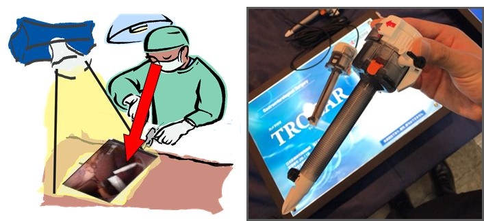
Surgical Navigation by Projection-Based Augmented Reality
In order to reduce the burden on the surgeon in laparoscopic surgery, we propose a surgical navigation system using augmented reality technology by projection. We also propose a novel medical imaging device named "trocar vision" that incorporates a small camera in a trocar's tube used in laparoscopic surgery and we are developing it for practical use. This enables laparoscopic surgery while observing inside the body cavity from a multidirectional viewpoint, and it is expected to improve safety and improve accuracy.
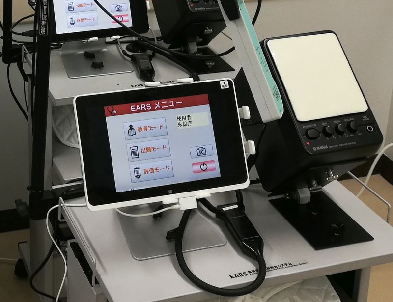
Augmented Reality Medical Training System
Currently, auscultation training in medical education is mainly used for mannequin type devices, but it has a cost problems such as expensive and large space requirement. Therefore, we propose an augmented reality auscultation training system named 'EARS' which reproduces arbitrary heart sound / respiratory sound by touching a virtual stethoscope onto a healthy person's body. This can reproduce various pathological conditions for effective auscultation training.
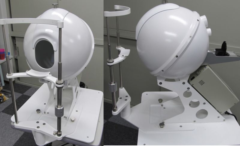
Computer-Aided Diagnosis
Although the tongue changes variously, reflecting the health condition of the whole body, objective recording methods are not established. Therefore we propose a tongue imaging system named 'TIAS' which quantitatively and stably records the tongue color, shape, wetness and so on. Beside that we established a database of tongue color and Kampo findings and constructed a diagnostic support system that estimates Kampo findings from a tongue picture using machine learning techniques. In addition, we are conducting research on cerebral aneurysm detection by automatic processing from MRA images, and automatic detection of lesion sites such as metastatic cancer and cysts that occurred in the liver from CT images.